APC Conjugated Anti-AGPS Monoclonal Antibody (Clone: AGPS-03)

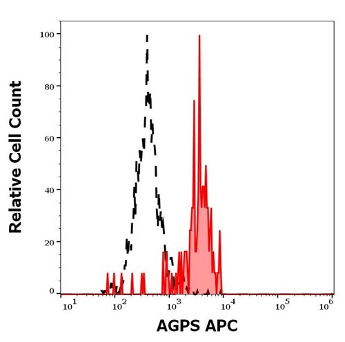
Figure 1: Separation of A431 cells stained using anti-AGPS (MHD4-46) APC antibody (10 μl reagent per million cells in 100 μl of cell suspension, red-filled) from A431 cells stained using mouse IgG2a isotype control (MOPC-173) APC antibody (concentration in sample 5 μg/ml, same as AGPS APC antibody concentration, black-dashed) in flow cytometry analysis (intracellular staining).
Roll over image to zoom in
Shipping Info:
For estimated delivery dates, please contact us at [email protected]
| Format : | Purified |
| Amount : | 0.1 mg |
| Isotype : | Mouse IgG2a |
| Purification : | Purified antibody is conjugated with activated allophycocyanin (APC) under optimum conditions and unconjugated antibody and free fluorochrome are removed by size-exclusion chromatography. |
| Content : | 0.1 mg/ml Stabilizing phosphate buffered saline (PBS), pH 7.4, 15 mM sodium azide |
| Storage condition : | Store at 2-8°C protected from light. Do not freeze. |
AGPS (alkylglycerone phosphate synthase), is an enzyme that catalyzes the second step of ether lipid biosynthesis in which acyl-dihydroxyacetone phosphate (acyl-DHAP) is converted to alkyl-DHAP by addition of a long chain alcohol and removal of a long-chain acid anion. The protein is localized to the inner side of the peroxisomal membrane and requires FAD as a cofactor. Mutations in AGPS gene have been associated with type 3 of rhizomelic chondrodysplasia punctata (RCDP3), and Zellweger syndrome. Higher expression of AGPS was observed in BCR/ABL positive leukemias and it was also described to be associated with higher risk of relapse.
Flow cytometry: The reagent is designed for analysis of human blood cells using 10 μl reagent / 100 μl of whole blood or 106 cells in a suspension. The content of a vial (1 ml) is sufficient for 100 tests. Intracellular staining.
For Research Use Only. Not for use in diagnostic/therapeutics procedures.
|
There are currently no product reviews
|


.png)


