PE-Cy7 Conjugated Anti-CD79a Monoclonal Antibody (Clone:HM47)

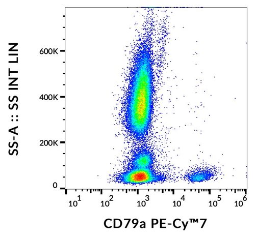
Figure 1: Flow cytometry analysis (intracellular staining) of CD79a in human peripheral blood with anti-CD79a (HM47) PE-Cy™7.
Roll over image to zoom in
Shipping Info:
For estimated delivery dates, please contact us at [email protected]
| Format : | Purified |
| Amount : | 100 Tests |
| Isotype : | Mouse IgG1 |
| Purification : | Purified antibody is conjugated with activated tandem dye of R-phycoerythrin-cyanine 7 (PE-Cy™7) under optimum conditions and unconjugated antibody and free fluorochrome are removed by size-exclusion chromatography. |
| Content : | Stabilizing phosphate buffered saline (PBS), pH 7.4, 15 mM sodium azide |
| Storage condition : | Store at 2-8°C protected from light. Do not freeze. |
CD79a (Ig alpha, MB1) forms disulfide-linked heterodimer with CD79b (Ig beta). They both are transmembrane proteins with extended cytoplasmic domains containing immunoreceptor tyrosine activation motives (ITAMs), and together with cell surface immunoglobulin they constitute B-cell antigen-specific receptor (BCR). CD79a and b are the first components of BCR that are expressed developmentally. They appear on pro-B cells in association with the endoplasmic reticulum chaperone calnexin. Subsequently, in pre-B cells, CD79 heterodimer is associated with lambda5-VpreB surrogate immunoglobulin and later with antigen-specific surface immunoglobulins. At the plasma cell stage, CD79a is present as an intracellular component. CD79a/b complex interacts with Src-family tyrosine kinase Lyn, which phosphorylates its cytoplasmic ITAM motives to form docking sites for downstream signaling.
Flow cytometry: The reagent is designed for analysis of human blood cells using 4 μl reagent / 100 μl of whole blood or 106 cells in a suspension. The content of a vial (0.4 ml) is sufficient for 100 tests. Intracellular staining.
For Research Use Only. Not for use in diagnostic/therapeutics procedures.
|
There are currently no product reviews
|


.png)


