Mouse Monoclonal Antibody To N-Cadherin (Clone: 8C11)

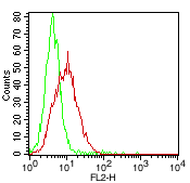
Fig. 1: Cell Surface FLOW analysis of N-Cadherin in KG1 cells using 0.5 µg of antibody (Clone: 8C11). Green represents isotype control; red represents anti-N Catherine (10-7653) antibody. Goat anti-mouse PE conjugate was used as secondary antibody.
Roll over image to zoom in
Shipping Info:
Order now and get it on Tuesday April 29, 2025
Same day delivery FREE on San Diego area orders placed by 1.00 PM
| Format : | Purified |
| Amount : | 100 µg |
| Isotype : | Mouse IgG1 kappa |
| Purification : | Affinity Chromatography |
| Content : | 0.5 mg/ml of Ab purified from Bioreactor Concentrate by Protein A/G. Prepared in 10 mM PBS with 0.05% BSA & 0.05% azide. |
| Storage condition : | Store the antibody at 4°C; stable for 6 months. For long-term storage; store at -20°C. Avoid repeated freeze and thaw cycles. |
N-cadherin(CD325) is a 140 kD protein belongs to a transmembrane molecules that mediate calcium dependent intracellular adhesion. Its extracellular region consists of five EC domains and has one cytoplasmic domain. N-cadherin is involved in organogenesis and maintenance of organ architecture by contributing to the sorting of heterogeneous cell types and in the cell adhesion needed to form tissues. N-cadherin is expressed by stem cells, myeloblasts, endothelial cells, and fibroblasts, and also is expressed in neural and muscle tissues and some types of carcinoma cells. CD325 associates with the cytoskeleton trough catenin proteins.
For Research Use Only. Not for use in diagnostic/therapeutics procedures.
| Subcellular location: | Cell membrane, Cell membrane, Cell junction, Cell surface |
| Post transnational modification: | May be phosphorylated by OBSCN. |
| BioGrid: | 107435. 61 interactions. |
|
There are currently no product reviews
|


.png)


