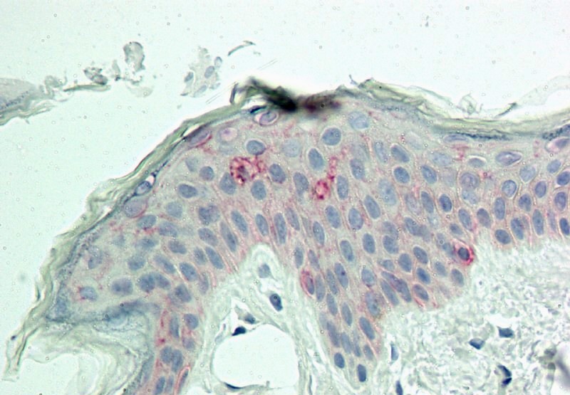
Monoclonal Antibody to MITF (Clone: ABM1H91)

Fig-1: Western blot analysis of MITF. Anti- MITF antibody (Clone: ABM1H91) was tested at 4 µg/ml on mouse Brain lysate.
Roll over image to zoom in
Shipping Info:
Order now and get it on Tuesday April 29, 2025
Same day delivery FREE on San Diego area orders placed by 1.00 PM
| Format : | Purified |
| Amount : | 100 µg |
| Isotype : | Mouse IgG1 Kappa |
| Purification : | Protein G Chromatography |
| Content : | 25 µg in 50 µl/100 µg in 200 µl PBS containing 0.05% BSA and 0.05% sodium azide. Sodium azide is highly toxic. |
| Storage condition : | Store the antibody at 4°C, stable for 6 months. For long-term storage, store at -20°C. Avoid repeated freeze and thaw cycles. |
MITF (Microphthalmia-associated transcription factor) is a basic helix-loop-helix leucine zipper (bHLH-Zip) family factor, involved in lineage-specific pathway regulation of many types of cells including osteoclasts, melanocytes, retinal pigment epithelial cells, mast cells and natural killer cells. It plays a critical role in melanogenesis as a transcriptional activator of tyrosinase, TRP-1, and TRP-2. Moreover, it regulates specification, survival, and proliferation of normal melanocytes, and controls proliferation, migration and invasion of melanoma cells. MITF is the most characterized member of the MIT family, mutations of which can lead to diseases such as Melanoma, Waardenburg syndrome, and Tietz syndrome.
Western blot analysis: 2-4 µg/ml, Immunohistochemical analysis: 10 µg/ml
For Research Use Only. Not for use in diagnostic/therapeutics procedures.
| Subcellular location: | Nucleus |
| Post transnational modification: | Ubiquitinated following phosphorylation at Ser-180, leading to subsequent degradation by the proteasome. Deubiquitinated by USP13, preventing its degradation. |
| Tissue Specificity: | Isoform M is exclusively expressed in melanocytes and melanoma cells. Isoform A and isoform H are widely expressed in many cell types including melanocytes and retinal pigment epithelium (RPE). Isoform C is expressed in many cell types including RPE but not in melanocyte-lineage cells. Isoform Mdel is widely expressed in melanocytes, melanoma cell lines and tissues, but almost undetectable in non-melanoma cell lines. |
| BioGrid: | 110432. 27 interactions. |
|
There are currently no product reviews
|


.png)


