APC Conjugated Anti-EGFR Monoclonal Antibody (Clone:EGFR1)

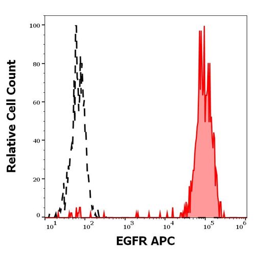
Figure 1: Separation of A431 cells stained using anti-EGFR (EGFR1) APC antibody (10 μl reagent per million cells in 100 μl of cell suspension, red-filled) from A431 cells stained using mouse IgG2b isotype control (MPC-11) APC antibody (concentration in sample 5 μg/ml, same as EGFR APC antibody concentration, black-dashed) in flow cytometry analysis (surface staining).
Roll over image to zoom in
Shipping Info:
For estimated delivery dates, please contact us at [email protected]
| Format : | Purified |
| Amount : | 100 Tests |
| Isotype : | Mouse IgG2b |
| Purification : | Purified antibody is conjugated with activated allophycocyanin (APC) under optimum conditions and unconjugated antibody and free fluorochrome are removed by size-exclusion chromatography. |
| Storage condition : | Store at 2-8°C protected from light. Do not freeze. |
The oncoprotein EGFR (epidermal growth factor receptor), also known as HER1 / ErbB1, is a member of ErbB family of cell surface receptor tyrosine kinases. This 170 kDa transmembrane glycoprotein is often associated with cancerogenesis, although its activation state is controlled at various levels including trafficking and degradation steps. Binding of receptor-specific ligands to the EGFR ectodomain results in formation of homodimeric or heterodimeric kinase-active complexes into which HER2 / ErbB2 is recruited as a preferred partner. Subsequent signaling cascades such as RAS/MAPK and PI3K/AKT pathways lead to cell proliferation and survival responses.
For Research Use Only. Not for use in diagnostic/therapeutics procedures.
|
There are currently no product reviews
|


.png)


