Anti-Vimentin Monoclonal Antibody (Clone:VI-RE/1)

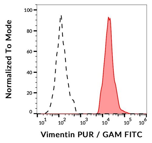
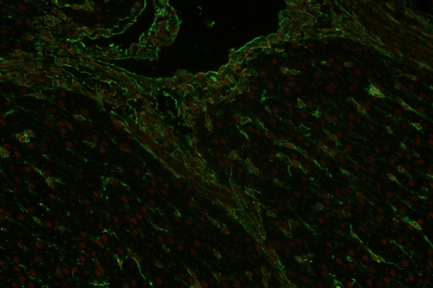
Figure 1: Flow cytometry analysis (intracellular staining) of vimentin expression in ESS-1 cells using anti-human vimentin (VI-RE/1) purified, GAM-FITC. Negative control: human lymphocytes
Roll over image to zoom in
Shipping Info:
For estimated delivery dates, please contact us at [email protected]
| Format : | Purified |
| Amount : | 0.1 mg |
| Isotype : | Mouse IgG1 |
| Purification : | Purified by protein-A affinity chromatography |
| Storage condition : | Store at 2-8°C. Do not freeze. |
Vimentin (57 kDa) is the most ubiquituos intermediate filament protein and the first to be expressed during cell differentiation. All primitive cell types express vimentin but in most non-mesenchymal cells it is replaced by other intermediate filament proteins during differentiation. Vimentin is expressed in a wide variety of mesenchymal cell types - fibroblasts, endothelial cells etc., and in a number of other cell types derived from mesoderm, e.g., mesothelium and ovarian granulosa cells. In non-vascular smooth muscle cellsand striated muscle, vimentin is often replaced by desmin, however, during regeneration, vimentin is reexpressed. Cells of the lymfo-haemopoietic system (lymphocytes, macrophages etc.) also express vimentin, sometimes in scarce amounts. Vimentin is also found in mesoderm derived epithelia, e.g. kidney (Bowman capsule), endometrium and ovary (surface epithelium), in myoepithelial cells (breast, salivary and sweat glands), an in thyroid gland epithelium. In these cell types, as in mesothelial cells, vimentin is coexpressed with cytokeratin. Furthermore, vimentin is detected in many cells from the neural crest. Particularly melanocytes express abundant vimentin. In glial cells vimentin is coexpressed with glial filament acidic protein (GFAP).Vimentin is present in many different neoplasms but is particulary expressed in those originated from mesenchymal cells. Sarcomas e.g., fibrosarcoma, malignt fibrous histiocytoma, angiosarcoma, and leio- and rhabdomyosarcoma, as well as lymphomas, malignant melanoma and schwannoma, are virtually always vimentin positive. Mesoderm derived carcinomas like renal cell carcinoma, adrenal cortical carcinoma and adenocarcinomas from endometrium and ovary usually express vimentin. Also thyroid carcinomas are vimentin positive. Any low differentiated carcinoma may express some vimentin. Vimentin is frequently included in the so-called primary panel (together with CD45, cytokeratin, and S-100 protein). Intense staining reaction for vimentin without coexpression of other intermediate filament proteins is strongly suggestive of a mesenchymal tumour or malignant melanoma.
Flow Cytometry Recommended dilution:1-10 µg/ml
Western Blotting Recommended dilution: 1-2 µg/ml, overnight in 4
For Research Use Only. Not for use in diagnostic/therapeutics procedures.
| Subcellular location: | Cytoplasm, Cytoplasm, Nucleus matrix |
| Post transnational modification: | S-nitrosylation is induced by interferon-gamma and oxidatively-modified low-densitity lipoprotein (LDL(ox)) possibly implicating the iNOS-S100A8/9 transnitrosylase complex. |
| BioGrid: | 113272. 291 interactions. |
|
There are currently no product reviews
|


.png)


