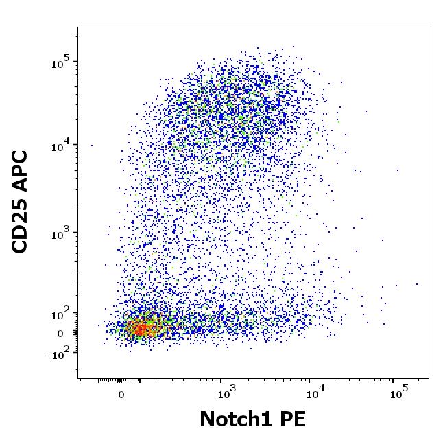
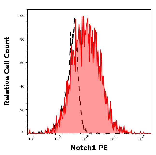
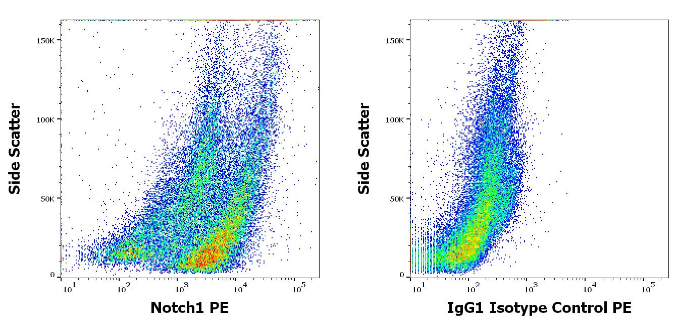
PE Conjugated Anti-Notch 1 Monoclonal Antibody (Clone:mN1A)

Figure 1: Intracellular staining of Notch1 in Jurkat cells using anti-Notch1 (mN1A) PE.
Roll over image to zoom in
Shipping Info:
For estimated delivery dates, please contact us at [email protected]
| Amount : | 0.1 mg |
| Isotype : | Mouse IgG1 |
| Content : | Antibody suspended in phosphate buffered saline (PBS) solution containing 15 mM sodium azide |
| Storage condition : | Store in the dark at 2-8°C. Do not freeze. Avoid prolonged exposure to light. |
Notch 1 is a 270-300 kDa transmembrane heterodimeric protein with multiple extracellular growth factor-like repeats, and with an intracellular domain consisting of multiple different domain types. It serves as a receptor for membrane ligands, such as Delta 1, Jagged1 (CD339), and Jagged2, and regulates cell fate decisions. Upon ligand binding the transmembrane form of Notch 1 is repeatedly cleaved to provide approximately 120 kDa Notch intracellular fragment (NICD), which translocates to the nucleus and acts as a part of transcriptional complexes that alter differentiation, proliferation, and apoptosis. The highest level of Notch 1 expression is in brain, lung and thymus.
| Subcellular location: | Nucleus |
| Post transnational modification: | Hydroxylated at Asn-1955 by HIF1AN. Hydroxylated at Asn-2022 by HIF1AN (By similarity). Hydroxylation reduces affinity for HI1AN and may thus indirectly modulate negative regulation of NICD (By similarity). |
| Tissue Specificity: | In fetal tissues most abundant in spleen, brain stem and lung. Also present in most adult tissues where it is found mainly in lymphoid tissues. |
| BioGrid: | 110913. 223 interactions. |
|
There are currently no product reviews
|


.png)


