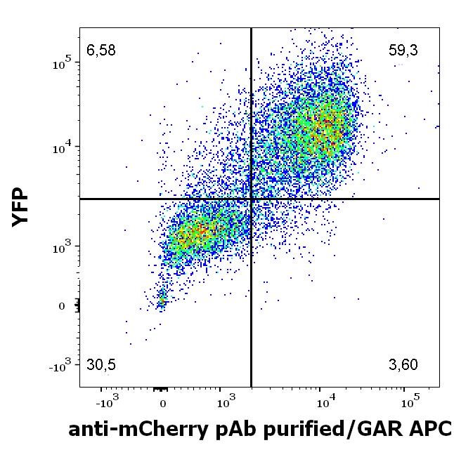
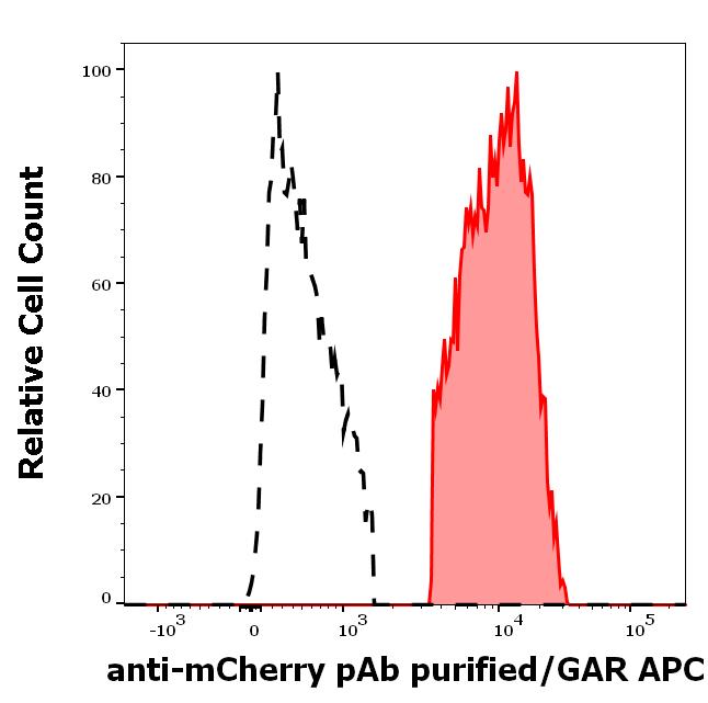
Anti-mCherry purified

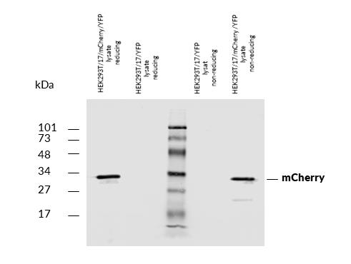
Figure 1: Western blotting analysis of mCherry fluorescent protein using rabbit polyclonal PAb (918) on lysates of HEK293T/17 cells co-transfected with mCherry/GPI and YFP/GPI constructs (HEK293T/17 cells transfected with YFP/GPI; negative control) under reducing and non-reducing conditions. Nitrocellulose membrane was probed with 2 µg/ml of rabbit anti-mCherry polyclonal antibody followed by IRDye800-conjugated anti-rabbit secondary antibody. A specific band was detected for mCherry protein at approximately 30 kDa.
Roll over image to zoom in
Shipping Info:
For estimated delivery dates, please contact us at [email protected]
| Format : | Purified |
| Amount : | 0.1 mg |
| Purification : | Purified by ligand affinity chromatography. |
| Content : | Concentration: 1 mg/ml Storage Buffer: Stabilizing phosphate buffered saline (PBS), pH 7.4, 15 mM sodium azide |
| Storage condition : | Store at 2-8°C. Do not freeze. |
| Gene : | mCherry |
| Uniprot ID : | X5DSL3 |
| Immunogen Information : | mCherry protein from Anaplasma marginale |
The mCherry is a red fluorescent protein with excitation maximum 587 nm and emission maximum 610 nm. It has around 28 kDa, and it is being used as a fluorescent tag in expression systems.
Specificity :The rabbit polyclonal antibody PAb (918) recognizes a red fluorescent protein tag mCherry.
Flow cytometry: Recommended dilution: 1-4 µg/ml, extracellular staining or intracellular staining - depending on expression.
Western blotting: Recommended dilution: 1-2 µg/ml.
For Research Use Only. Not for use in diagnostic/therapeutics procedures.
|
There are currently no product reviews
|


.png)


