Anti-Cytokeratin 8 Monoclonal Antibody (Clone:C-43)-FITC Conjugated

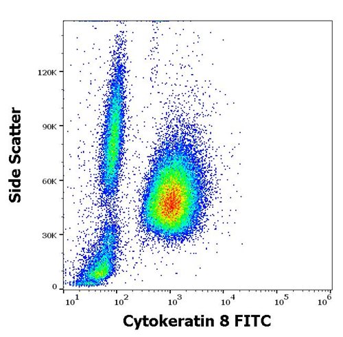
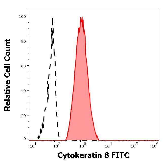
Figure 1: Flow cytometry intracellular staining pattern of human peripheral whole blood mixed with A431 cellular suspension stained using anti-Cytokeratin 8 (C-43) FITC antibody
Roll over image to zoom in
Shipping Info:
For estimated delivery dates, please contact us at [email protected]
| Amount : | 0.1 mg |
| Isotype : | Mouse IgG1 |
| Storage condition : | Store in the dark at 2-8°C. Do not freeze. Avoid prolonged exposure to light. |
Cytokeratins are a subfamily of intermediate filaments and characterized by remarkable biochemical diversity. Cytokeratins are represented in epithelial tissues by at least 20 different polypeptides, molecular weight between 40 kDa and 68 kDa. The individual cytokeratin polypeptides are designated 1 to 20 and divided into the type I (acidic cytokeratins 9-20) and type II (basic to neutral cytokeratins 1-8) families.
| Subcellular location: | Cytoplasm, Nucleus, Nucleus matrix |
| Post transnational modification: | O-glycosylated (O-GlcNAcylated), in a cell cycle-dependent manner. |
| Tissue Specificity: | Observed in muscle fibers accumulating in the costameres of myoplasm at the sarcolemma membrane in structures that contain dystrophin and spectrin. Expressed in gingival mucosa and hard palate of the oral cavity. |
| BioGrid: | 110054. 69 interactions. |
|
There are currently no product reviews
|


.png)


