Anti-CD1c Monoclonal Antibody (Clone:L161)-PE Conjugated

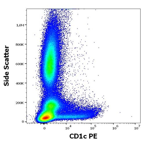
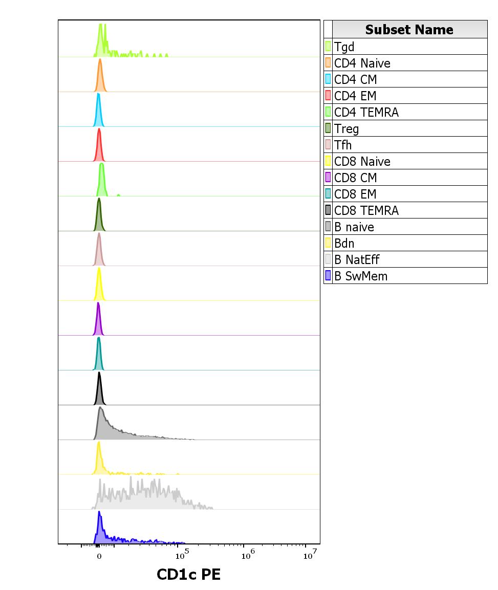
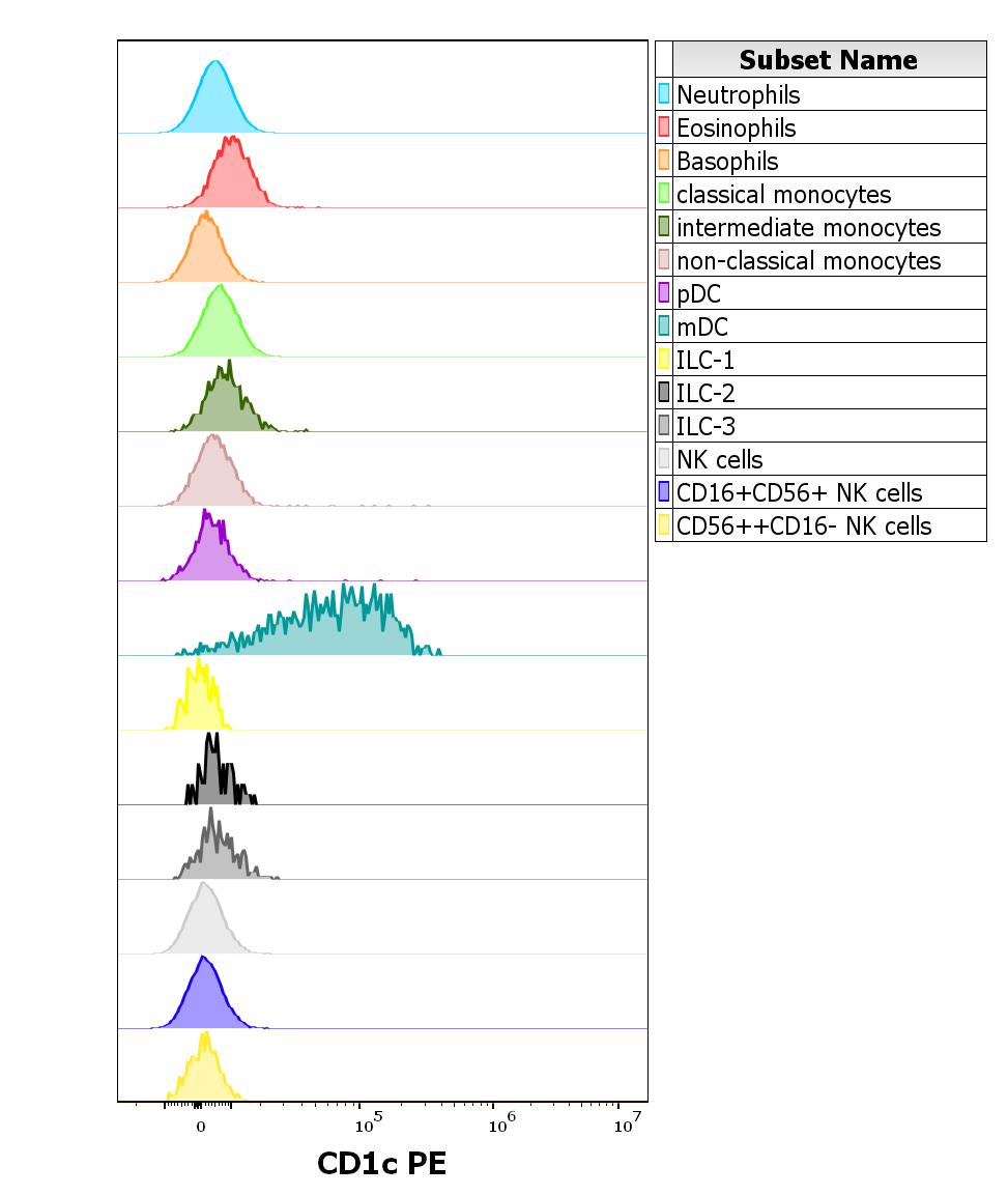
Figure 1: Analysis of the antibody staining profile was performed on blood leukocytes isolated from buffy coats. HCDM CDMaps standardized procedures were used for cell isolation and surface staining of blood leukocytes, with the modification of staining protocol using cytometry test tubes.Mouse monoclonal anti-human CD1c PE antibody (clone L161) was used in concentration 1 µg/ml in stained blood sample (2 x 10⁶ cells).
Roll over image to zoom in
Shipping Info:
For estimated delivery dates, please contact us at [email protected]
| Amount : | 100 tests |
| Isotype : | Mouse IgG1 |
| Storage condition : | Store in the dark at 2-8°C. Do not freeze. Avoid prolonged exposure to light. |
CD1c (also known as R7 or BDCA1) together with CD1a and b, belongs to group 1 of CD1 antigens. These non-classical MHC-like glycoproteins serve as antigen-presenting molecules for a subset of T cells that responds to specific lipids and glycolipids found in the cell walls of bacterial pathogens or self-glycolipid antigens such as gangliosides, and they have also roles in antiviral immunity. The trafficking routes of the particular CD1 types differ and correspond to their ability to bind and present different groups of antigens. CD1c is unique in its ability to present e.g. mycobacterial phosphoketides and polyisoprenoids. CD1c is the only CD1 isoform that has been shown to interact both with alpha/beta and gamma/delta T cells.
| Subcellular location: | Cell membrane, Endosome membrane, Lysosome |
| Tissue Specificity: | Expressed on cortical thymocytes, on certain T-cell leukemias, and in various other tissues. |
|
There are currently no product reviews
|


.png)


