Anti-CD146 Monoclonal Antibody (Clone:P1H12)-PE Conjugated

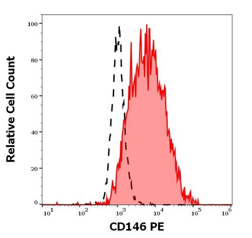
Figure 1: Separation of cell stained using anti- h CD146 PE antibody from cell stained using mouse IgG1 isotype control in flow cytometry analysis of HUVEC cell suspension
Roll over image to zoom in
Shipping Info:
For estimated delivery dates, please contact us at [email protected]
| Amount : | 100 tests |
| Isotype : | Mouse IgG1 |
| Storage condition : | Store in the dark at 2-8°C. Do not freeze. Avoid prolonged exposure to light. |
CD146, also known as MCAM (melanoma cell adhesion molecule) or MUC18, is a heavily glycosylated transmembrane glycoprotein with more than 50% of the mass from carbohydrates. It is expressed on epithelial and endothelial cells, fibroblasts, multipotent mesenchymal stromal cells, activated T cells and activated keratinocytes, and on some cancer cells, especially melanoma. The presence of CD146 on circulating blood cells has been confined to the activated T cells rather than circulating endothelial cells. CD146 mediates heterophilic cell adhesion and regulates monocyte transendothelial migration.
Flow cytometry: The reagent is designed for analysis of human blood cells using 10 μl reagent / 100 μl of whole blood or 106 cells in a suspension. The content of a vial (1 ml) is sufficient for 100 tests.
For Research Use Only. Not for use in diagnostic/therapeutics procedures.
| Subcellular location: | Membrane |
| Tissue Specificity: | Detected in endothelial cells in vascular tissue throughout the body. May appear at the surface of neural crest cells during their embryonic migration. Appears to be limited to vascular smooth muscle in normal adult tissues. Associated with tumor progression and the development of metastasis in human malignant melanoma. Expressed most strongly on metastatic lesions and advanced primary tumors and is only rarely detected in benign melanocytic nevi and thin primary melanomas with a low probability of metastasis. |
| BioGrid: | 110332. 15 interactions. |
|
There are currently no product reviews
|


.png)


